Environmental factors, especially intestinal microbes and their toxins, may contribute to the development of autoimmune diseases such as rheumatoid arthritis (RA) (1-7), inflammatory bowel diseases (IBDs) (8, 9), systemic lupus erythematosis (SLE) (10), and other chronic disorders (11-14). Moreover, the translocation of live bacteria, their cellular components and toxins into the body is significantly increased by physical and psychological stress, drugs, surgery, and irregularity (15-17).
The immune system plays a key role in protecting the host against hazardous foreign substances. Moreover, a properly functioning immune system is also affected by a variety of exogenous environmental factors (infectious bacteria and viruses), drugs (immunosuppressants and immunostimulants), and intrinsic factors (genetics, stress, gastrointestinal disorders, and immunosenescence associated with aging). In fact, lowered immune function may increase health risks, such as susceptibility to disease causative pathogens. Because immune function can be evaluated by determining the antibody response to foreign antigens, Chondrex, Inc. provides ELISAs to measure IgA and IgG antibody levels to various types of pathogenic and non-pathogenic environmental factors. While these antibody ELISA kits against environmental factors are useful as immune function evaluation tools, several considerations must be addressed before establishing study protocols. Here we would like to share these considerations to help users better choose kits and interpret results. Please contact us for more information at support@chondrex.com.
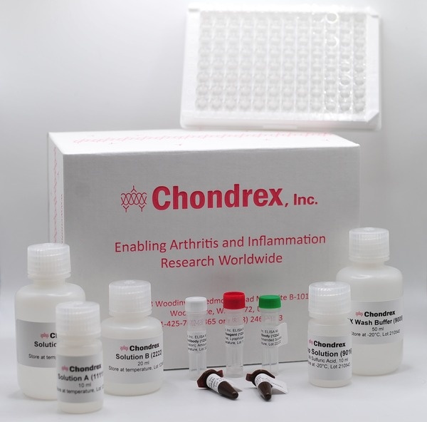
This series of ELISA kits employ the ChonBlockTM buffer system, which eliminates non-specific hydrophobic and ionic interactions in indirect ELISAs to provide accurate assay results. Learn more about how ChonBlock can improve your assay results!
Human Anti-Bacteria/Toxin IgA Antibody Assay Kits

Human Anti-Bacteria/Toxin IgG Antibody Assay Kits
Background
Lipopolysaccharide (LPS) is the dominant bacterial toxin in the intestinal tract and plays important roles in the development of the host's immune system. However, it is also highly likely that LPS plays important pathological roles in patients with chronic autoimmune diseases.
LPS is known to polyclonally active B-cells and consequently activate T-cells, but also triggers and exacerbates inflammatory reactions (5, 6), and may be involved in a pathogenic nature in various chronic diseases such as diabetes (14), systemic lupus erythematosus (10), and autoimmune hemolytic anemia (11). Therefore, in order to understand the possible involvement of these environmental factors in the pathogenesis of autoimmune diseases, it is important to distinguish how healthy people and patients with diseases respond to these common bacterial toxins.

Pathogenic Environmental Factors and the Possible Contribution to Autoimmune Diseases
Various factors can disrupt the mucosal barrier function (a). As a result, pathogenic intestinal bacterial components (mimic antigens) and their toxins can cross the mucosal barrier into the surrounding tissues and circulation, thus disrupting the immune homeostasis (b).
References
- Aoki S, Y.K., Yokoyama T, Nonogaki T, Iwasaki S, Mitsui T, Niwa S., Role of enteric bacteria in the pathogenesis of rheumatoid arthritis: evidence for antibodies to enterobacterial common antigens in rheumatoid sera and synovial fluids. Ann Rheum Dis., 1996. 55: 363-9.
- Van der Heijden IM, W.B., Tchetverikov I, Schrijver IA, Schouls LM, Hazenberg MP, Breedveld FC, Tak PP. Presence of bacterial DNA and bacterial peptidoglycans in joints of patients with rheumatoid arthritis and other arthritides. Arthritis Rheum., 2000. 43:593-8.
- Terato K, Y.X., Miyahara H, Cremer MA, Griffiths MM., Induction of chronic autoimmune arthritis in DBA/1 mice by oral administration of type II collagen and E.coli LPS. Br J Rheum, 1996. 35: 828-838.
- Peltonen R, N.M., Helve T, Hänninen O, Toivanen P, Eerola E. Faecal microbial flora and disease activity in rheumatoid arthritis during a vegan diet. Br J Rheumatol, 1997. 36: 64-8.
- Toivanen, P., Normal intestinal microbiota in the aetiopathogenesis of rheumatoid arthritis. Ann Rheum Dis., 2003. 62: 807-11.
- Vaahtovuo J, M.E., Korkeamäki M, Luukkainen R, Toivanen P. Fecal microbiota in early rheumatoid arthritis. J Rheumatol., 2008. 35: 1500-5.
- Edwards, C., Commensal Gut Bacteria and the Etiopathogenesis of Rheumatoid Arthritis. J Rheum, 2008. 35: 1477-78.
- Dayna Shi, J.D., G.D., Inflammatory bowel disease requires the interplay between innate and adaptive immune signals. Cell Research, 2006. 16: 70-74.
- Nell S, S.S., Josenhans C, The impact of the microbiota on the pathogenesis of IBD: lessons from mouse infection models. Nat Rev Microbiol. 2010. 8: 564-77.
- Cavallo T, G.N., Bacterial lipopolysaccharide induces long-lasting IgA deficiency concurrently with features of polyclonal B cell activation in normal and in lupus-prone mice. Clin Exp Immunol, 1991. 84: 134-138.
- Murakami M, N.K., Yamazaki K, Muraguchi T, Serikawa T, Honjo T., Effects of breeding environments on generation and activation of autoreactive B-1 cells in anti-red blood cell autoantibody transgenic mice. Exp Med, 1997. 185: 791-4.
- Penhale WJ, Y.P., The influence of the normal microbial flora on the susceptibility of rats to experimental autoimmune thyroiditis. Clin Exp Immunol, 1988. 72: 288-92.
- Murakami M, T.T., Shinkura R, Nisitani S, Okamoto M, Yoshioka H, Usui T, Miyawaki S, Honjo T., Oral administration of lipopolysaccharides activates B-1 cells in the peritoneal cavity and lamina propria of the gut and induces autoimmune symptoms in an autoantibody transgenic mouse. J Exp Med, 1994. 180: 111-21.
- Nymark M, P.P., Tuomainen AM, Forsblom C, Groop PH, Lehto M; FinnDiane Study Group., Serum lipopolysaccharide activity is associated with the progression of kidney disease in Finnish patients with type 1 diabetes. Diabetes Care, 2009. 32: 1689-93.
- Anderlik P, S.I., Bános Z, Barna Z., Bacterial translocation after cold stress in young and old mice. Acta Microbiol Hung, 1990. 37: 289-94.
- Velin AK, E.A., Braaf Y, Wallon C, Söderholm JD., Increased antigen and bacterial uptake in follicle associated epithelium induced by chronic psychological stress in rats. Gut, 2004. 53: 494-500.
- Khalif IL, Q.E., Konovitch EA, Maximova ID., Alterations in the colonic flora and intestinal permeability and evidence of immune activation in chronic constipation. Dig Liver Dis, 2005. 37: 838-49.