Ovalbumin (OVA), consisting of 385 amino acid residues and a molecular weight of 43 kDa, has been used as an antigen for a diverse array of immunology studies due to its antigenicity and (typically) benign immune response. OVA has been used to induce IgE-mediated diseases, to evaluate vaccine delivery methods, and even to study tumor biology by using transgenic tumor cells expressing OVA as a tumor antigen. As such, OVA is a well-characterized and affordable antigen to study immune responses in a variety of contexts.
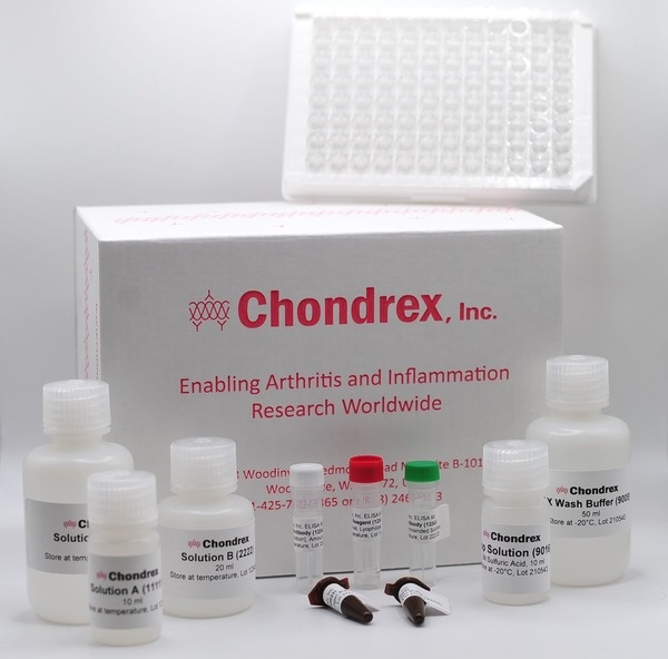
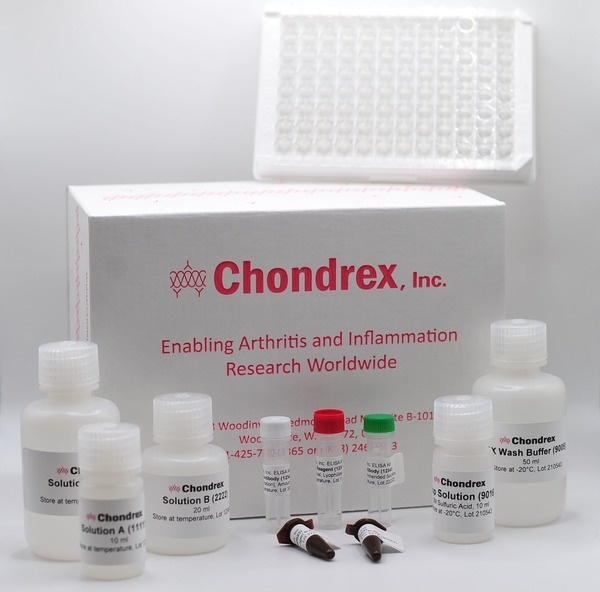
Chondrex, Inc.'s well-referenced, convenient, and easy-to-use Mouse Anti-OVA Antibody ELISA Kits can quantify OVA-specific isotypes and IgG subclasses of mouse antibodies, including: IgE*, IgG (IgG1, IgG2a, IgG2b, IgG2c), IgM, and IgA antibodies. To find references using these products, please click on the links in the table below. To learn more about each of our Mouse Anti-OVA Antibody ELISA Kits, continue reading below. If you need assistance in choosing the correct kit for your purposes, please contact us.
*When assaying for anti-OVA antibodies, especially IgE antibodies, total immunoglobulin assays are important tools to evaluate and normalize immune responses as a ratio of anti-OVA antibody levels to total antibody subtype levels.
ANNOUNCEMENT: Starting 10/01/2024, Chondrex, Inc.'s OVA Antibody Subtype Kits (Cat # 3011, 3013, 3015, 3016, 3017, 3018, and 3029) now use a new and improved OVA (Cat # 300411) for better plate coating. Customers who purchased any of the above kits which used OVA Cat # 30112 before 9/30/2024 , please refer to this protocol for plate coating instructions. All OVA antibody subtype kits purchased before 9/30/24 will still be valid through their provided expiration dates.
Mouse IgE and Anti-OVA Antibody Assay Kits
| Product | Catalog # | Price (USD) | |
|---|---|---|---|
 |
Mouse Anti-OVA IgA Antibody Assay Kit | 3018 | 439.00 |
 |
Mouse Anti-OVA IgE Antibody Assay Kit | 3004 | 444.00 |
 |
Mouse Anti-OVA IgG Antibody Assay Kit | 3011 | 439.00 |
 |
Mouse Anti-OVA IgG1 Antibody Assay Kit | 3013 | 439.00 |
 |
Mouse Anti-OVA IgG2a Antibody Assay Kit | 3015 | 439.00 |
 |
Mouse Anti-OVA IgG2b Antibody Assay Kit | 3016 | 439.00 |
 |
Mouse Anti-OVA IgG2c Antibody Assay Kit | 3029 | 439.00 |
 |
Mouse Anti-OVA IgM Antibody Assay Kit | 3017 | 439.00 |
 |
Mouse Serum Anti-OVA IgE Antibody Assay Kit | 3010 | 439.00 |
 |
Mouse Total IgA Antibody Detection Kit | 3019 | 355.00 |
 |
Mouse Total IgE (IgEa and IgEb) Detection Kit | 3005 | 444.00 |
Adjuvant Influence on Antibody Isotype Production
Choosing an appropriate adjuvant and immunization protocol is very important to induce the desired immune response to OVA. The adjuvant used can influence the antibody isotype produced. Aluminum adjuvants, widely used in the OVA-Induced Asthma model, can elicit anti-allergen IgE antibodies (1). Cholera toxins, with oral immunization, are effective at inducing IgA antibodies (2), while Complete Freund's Adjuvant (CFA) is commonly used to stimulate IgG and IgM production (3). These adjuvants shift the balance of Th1 and Th2 responses to OVA or other antigens, which subsequently influences immunoglobulin isotype production (4,5). When choosing an anti-OVA Antibody ELISA Kit to evaluate the efficacy of OVA sensitization/challenge models in animals, consider the adjuvant used in your OVA immunization protocol.
Selecting the Correct Antibody Isotype/Subtype Assay
Several other considerations must be made when choosing the appropriate Mouse Anti-OVA Antibody ELISA Kit for your experimental purposes. Below is key information that can aide in determining which anti-OVA antibody isotypes are relevant for your research goals.
When assaying for anti-OVA antibodies, especially IgE antibodies, it is important to evaluate and normalize the immune response to OVA by determining the ratio of anti-OVA antibody levels to total antibody subtype levels. For this purpose, Chondrex, Inc. provides Mouse Total Immunoglobulin Detection ELISA Kits.
Mouse Anti-OVA IgG (& IgG subclass) Antibody ELISA Kits
Immunoglobulin G (IgG) antibodies are the predominant antibody isotype found in blood and extracellular fluid. They possess the longest half-life of any antibody and have a relatively strong affinity for their target antigens. IgG antibodies serve several important functions in the immune response: neutralization of pathogens, opsonization of pathogens (marking pathogens for degradation), and complement fixation/activation. Each of the 4 known mouse subtypes of IgG (IgG1, IgG2a/c*, IgG2b, IgG3) have different properties and contributions to these functions (6). For instance, mouse IgG1 antibodies are not potent activators of complement cascade, but mouse IgG2a and IgG2b antibodies strongly activate complement. The Th1/Th2 balance of molecular signaling also influences IgG subtype expression: Th1 signaling stimulates IgG2a/c, IgG2b, and IgG3 production, while Th2 signaling stimulates IgG1 and IgE production (7,8).
Generally, quantifying mouse anti-OVA IgG antibodies is an important metric in almost all immunological studies in which OVA is used as a model antigen. In IgE mediated disease/hypersensitivity reaction research, IgG antibodies may develop immune complexes with allergens that aide in mast cell degranulation and initiation of the hypersensitivity reactions. For studies involving food allergies or mucosal immunity, anti-OVA IgG and anti-OVA IgA antibody levels can be used to evaluate intestinal mucosal barrier function (9). When OVA is used in transgenic cell lines as a pseudo-tumor antigen, anti-OVA IgG antibodies levels can help analyze the efficacy of cancer vaccines to produce anti-tumor responses (10,11). The Mouse Anti-OVA IgG Antibody ELISA Kits (IgG: cat# 3011, IgG1: cat# 3013, IgG2a: cat# 3015, IgG2b: cat#3016, IgG2c: cat# 3029) provide a convenient and cost-effective method to evaluate humoral immune responses to OVA.
*The occurrence of IgG2a or IgG2c antibodies depends on the genetic background of mice. For instance, Balb/c mice produce IgG2a while C57BL/c mice produce IgG2c antibodies.
Mouse Serum Anti-OVA IgE Antibody ELISA Kit
Immunoglobulin E (IgE) antibodies are usually concentrated around mast cells located just underneath the skin and mucosal barriers (intestinal tract, respiratory tract). In serum, IgE antibodies are generally present in 1/1000 the levels compared of IgG antibodies in serum. As key mediators in acute allergic responses, IgE antibodies will bind to Fc-receptors (FcεRI) on the surface of mast cells. These mast cell-associated IgE antibodies will form immunocomplexes with their specific antigens and form allergen bridges (cross-links) between FcεRI proteins. The accumulation of bridged IgE-FcεRI complexes induces mast cell degranulation and histamine release, leading to hypersensitivity responses. Early-phase hypersensitivity reactions occur within minutes of allergen exposure and are mediated not only by molecules like histamine, but also serine proteases, as well as cytokines like Tumor Necrosis Factor alpha (TNFα) and Vascular Endothelial Growth Factor (VEGF) (12). The late-phase allergic responses occur 2-9 hours after allergen exposure and are mediated by Th2 cells and their effector cytokines (Interleukin-4 (IL-4), IL-13, and IL-5) among other signaling molecules (13).
The Mouse Serum Anti-OVA IgE Antibody ELISA Kit (cat# 3010) is ideal for studies where OVA is used to induce mouse IgE-mediated allergic disease models such as atopic dermatitis, allergic asthma*, and food allergies. For this usage, it may also be beneficial to measure the total IgE level in samples using our Mouse Total IgE Detection ELISA Kit. Both the OVA specific and total IgE ELISA Kits will detect mouse IgEa (found in Balb/c mice) and IgEb (C57BL/6 mice) antibodies. Furthermore, measuring Th2-associated cytokine levels can provide deeper insight into the progression of OVA-induced allergic diseases.
Despite serum IgE levels being very low in serum (μg/ml levels), serum IgE antibody assays using an indirect ELISA format with allergen (OVA) coated plates will, generally, underestimate anti-allergen (OVA) IgE levels. Immunization of OVA (or other allergens) in mice will typically elicit anti-OVA IgG antibodies in mg/ml levels, much larger than the IgE levels in serum. In indirect ELISA systems, anti-OVA IgE and anti-OVA IgG antibodies in serum samples will compete to bind OVA coated on the ELISA plate, leading to an underestimation of IgE antibodies. To overcome this, the Mouse Serum Anti-OVA IgE Antibody ELISA Kit (sandwich ELISA) employs an anti-IgE monoclonal antibody coated plate, instead of an OVA coated plate. Please see the kit protocol for more information.
The Mouse Anti-OVA IgE Antibody ELISA Kit (cat# 3004, indirect ELISA) works with samples containing IgE antibody only, such as culture medium and purified IgE, as well as serum samples which received Protein A or G filtration for removing IgG antibodies.
*Chondrex, Inc. also offers Mouse Anti-House Dust Mite Antibody ELISA Kits that are ideal for evaluating the House Dust Mite Induced Asthma models of acute and chronic allergic asthma.
Mouse Anti-OVA IgA Antibody ELISA Kit
Immunoglobulin As (IgA) are the second most abundant antibodies in serum, where they are typically found in their monomeric form. At mucosal surfaces, secretory IgA antibodies are found as dimers with two IgA monomers linked by a joining chain (14). These mucosal antibodies play an essential role in preventing the invasion of pathogens and other foreign antigenic substances.
Recently, intestinal bacteria and the intestinal microbiome has been increasingly implicated in the pathogenesis of many diseases (15). Animal studies using oral administration of bacteria have been used to study this relationship. In these studies, secretory IgA antibody levels serve to evaluate mucosal immunity. For instance, intestinal permeability studies using administration of transgenic E. coli expressing OVA can use serum anti-OVA IgA antibody and fecal anti-OVA IgA antibody levels to evaluate mucosal immune response to OVA.*
The Mouse Anti-OVA IgA Antibody ELISA Kits, when used concomitantly with Mouse Anti-OVA IgG Antibody ELISA Kits, provide an easy way to evaluate mucosal barrier function for studies using OVA as an antigen.
*Genetic background can influence polyreactive IgA antibody levels in mice (16). Please consider the mouse strain used in your study when choosing a suitable kit.
Mouse Anti-OVA IgM Antibody ELISA Kit
Immunoglobulin M (IgM) is the first antibody isotype elicited in the humoral immune response because antibody class switching is not required for its production. These antibodies generally have low affinity for their antigens. However, IgM molecules form pentamers with ten antigen-binding sites, making them potent activators of the complement cascade. The rapid production of IgM antibodies makes them important players in preventing and limiting infections. Assaying for anti-OVA IgM antibodies functions as a reliable way to evaluate vaccination protocols utilizing OVA as an antigen (17).
In allergy studies, eliciting oral tolerance against an allergen is an important feature of immunotherapies. To evaluate the immunomodulator capabilities of experimental immunotherapies, anti-OVA IgG and anti-OVA IgM antibodies are important metrics to evaluate and compare initial and late-stage immune responses to OVA. IgM antibody levels are markers for the initial immune response, while IgG, IgA, and IgE are more indicative of late-stage immune response to OVA.
The Mouse Anti-OVA IgM Antibody ELISA Kit (cat# 3017) is ideal for evaluating early humoral responses to OVA in immunotherapy and vaccine research.
References
- N. Mizutani, H. Goshima, T. Nabe, S. Yoshino, Establishment and characterization of a murine model for allergic asthma using allergen-specific IgE monoclonal antibody to study pathological roles of IgE. Immunol Lett 141, 235-245 (2012).
- A. K. Gloudemans et al., The mucosal adjuvant cholera toxin B instructs non-mucosal dendritic cells to promote IgA production via retinoic acid and TGF-β. PLoS One 8, e59822 (2013).
- J. H. Kim, Y. K. Ahn, Effects of diphenyl dimethyl dicarboxylate on oral tolerance to ovalbumin in mice. J Toxicol Sci 20, 375-382 (1995).
- C. Kaplan et al., Th1 and Th2 cytokines regulate proteoglycan-specific autoantibody isotypes and arthritis. Arthritis Res 4, 54-58 (2002).
- S. R. Holdsworth, A. R. Kitching, P. G. Tipping, Th1 and Th2 T helper cell subsets affect patterns of injury and outcomes in glomerulonephritis. Kidney Int 55, 1198-1216 (1999).
- S. S. Weber, J. Ducry, A. Oxenius, Dissecting the contribution of IgG subclasses in restricting airway infection with Legionella pneumophila. J Immunol 193, 4053-4059 (2014).
- A. K. Abbas, K. M. Murphy, A. Sher, Functional diversity of helper T lymphocytes. Nature 383, 787-793 (1996).
- R. L. Coffman, D. A. Lebman, P. Rothman, Mechanism and regulation of immunoglobulin isotype switching. Adv Immunol 54, 229-270 (1993).
- Y. Hagiwara et al., Protective mucosal immunity in aging is associated with functional CD4+ T cells in nasopharyngeal-associated lymphoreticular tissue. J Immunol 170, 1754-1762 (2003).
- Y. Dölen et al., Nanovaccine administration route is critical to obtain pertinent iNKt cell help for robust anti-tumor T and B cell responses. OncoImmunology 9, 1738813 (2020).
- C. Cekic, Y.J. Day, D. Sag, J. Linden, Myeloid expression of adenosine A2A receptor suppresses T and NK cell responses in the solid tumor microenvironment. Cancer Res 74, 7250-7259 (2014).
- S. J. Galli, M. Tsai, A. M. Piliponsky, The development of allergic inflammation. Nature 454, 445-454 (2008).
- S. J. Galli, M. Tsai, IgE and mast cells in allergic disease. Nat Med 18, 693-704 (2012).
- J. M. Woof, M. A. Kerr, The function of immunoglobulin A in immunity. J Pathol 208, 270-282 (2006).
- J. Durack, S. V. Lynch, The gut microbiome: Relationships with disease and opportunities for therapy. J Exp Med 216, 20-40 (2019).
- F. Fransen et al., BALB/c and C57BL/6 Mice Differ in Polyreactive IgA Abundance, which Impacts the Generation of Antigen-Specific IgA and Microbiota Diversity. Immunity 43, 527-540 (2015).
- J. H. Kim, M. Ohsawa, Oral tolerance to ovalbumin in mice as a model for detecting modulators of the immunologic tolerance to a specific antigen. Biol Pharm Bull 18, 854-858 (1995).