Goodpasture syndrome, an autoimmune disease caused by autoantibodies against the glomerular basement membrane (GBM) in the kidney and lung, manifests as severe renal disease and pulmonary hemorrhage. Patient autoantibodies recognize the non-collagenous domain of the alpha chains (α1 - α6) of type IV collagen (NC1), which comprises the GBM. More specifically, the primary Goodpasture antigen was identified in humans as the NC1 domain of the α3 chain of type IV collagen [α3(IV)NC1] (1-3) and was confirmed in several animal models (3, 4). Furthermore, Kohda et al. reported that the disease can be induced in Wistar Kyoto (WKY) rats by immunizing them with recombinant α4(IV)NC1 fragments, or administering anti-α4(IV)NC1 antibodies (5).
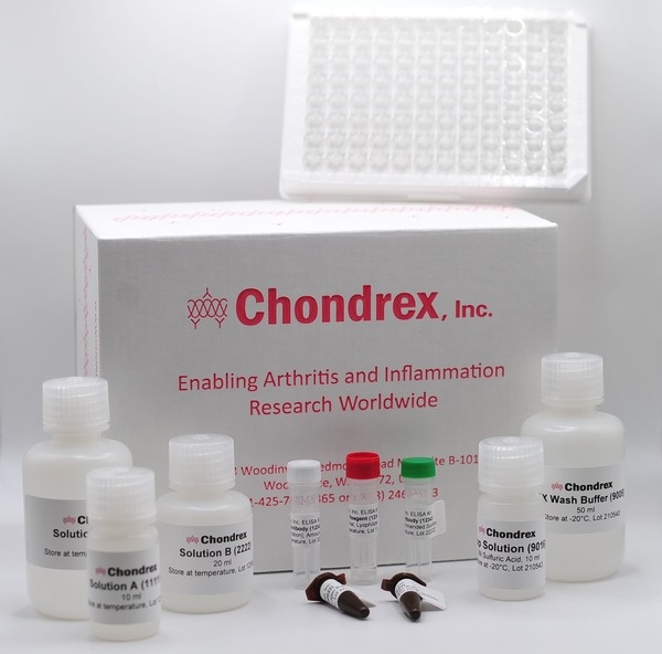
To induce rat experimental autoimmune glomerulonephritis, Chondrex, Inc. provides anti-NC1 antibodies and NC1 fragments. Rat anti-α4(IV)NC1 monoclonal antibodies can induce nephritis with a single intraperitoneal (IP) or intravenous (IV) injection, providing a Masugi nephritis model (6) (See Rat Nephritogenic Monoclonal Antibodies to Glomerular Basement Membrane Protocol). Immunizing with bovine NC1 fragments of type IV collagen provides a model for Steblay nephritis (7, 8) (See Experiment Autoimmune Glomerulonephritis Protocol). Chondrex, Inc. also offers mouse and rat renal NC1 fragments as an ELISA antigen to quantify cross-reactivity of autoantibodies to immunized bovine NC1 fragments.
The following information about these models is intended to help you understand glomerulonephritis and choose an appropriate disease model for your experiments. If you have any questions about this model, please contact support@chondrex.com for more information.
Inducing Rat Anti-GBM Nephritis Using Nephritogenic Antigens
| Product | Catalog # | Price (USD) | |
|---|---|---|---|
 |
Complete Freund's Adjuvant, 1 mg/ml | 7008 | 46.00 |
 |
Incomplete Freund's Adjuvant | 7002 | 40.00 |
 |
NC1 Fragment of Bovine GBM Type IV Collagen | 1102 | 342.00 |
 |
NC1 Fragment of Mouse Renal Type IV Collagen | 1106 | 378.00 |
 |
NC1 Fragment of Rat Renal Type IV Collagen | 1104 | 378.00 |
Inducing Rat Anti-GBM Nephritis Using Nephritogenic Monoclonal Antibodies
| Product | Catalog # | Price (USD) | |
|---|---|---|---|
 |
Nephritogenic Monoclonal Antibody, Clone 114, 1 mg | 70201 | 761.00 |
 |
Nephritogenic Monoclonal Antibody, Clone 114, 5 mg | 70205 | 3699.00 |
 |
Nephritogenic Monoclonal Antibody, Clone a84, 1 mg | 70211 | 761.00 |
 |
Nephritogenic Monoclonal Antibody, Clone a84, 5 mg | 70215 | 3699.00 |
 |
Nephritogenic Monoclonal Antibody, Clone b35, 1 mg | 70221 | 761.00 |
 |
Nephritogenic Monoclonal Antibody, Clone b35, 5 mg | 70225 | 3699.00 |
Rat Assays For Evaluating Disease Progress
| Product | Catalog # | Price (USD) | |
|---|---|---|---|
 |
Rat Albumin Detection Kit | 3020 | 355.00 |
 |
Rat Urinary Protein Assay Kit | 9040 | 115.00 |
 |
Rat Urinary Protein Assay Standard | 90401 | 65.00 |
Chondrex, Inc. also offers cationic bovine serum albumin to induce Immune Complex Glomerulonephritis in mice. Please refer to the animal model menu for more information.
Table of Contents
- Experimental Principle
- Protocol and Results
- Factors to Consider
- Background of Disease Models
1. Principle of Experimental Anti-Glomerular Basement Membrane Disease
Goodpasture disease, which is an autoimmune disease caused by anti-GBM antibodies, is characterized by glomerulonephritis and pulmonary hemorrhage. The GBM consists of trimers of alpha chains (α1 - α6) of type IV collagen. Among the 6 alpha chains, the non-collagenous domain 1 of the alpha 3 chain of type IV collagen (α3(IV)NC1) was reported as the primary antigen in human Goodpasture disease (9).
In order to study autoimmune-mediated nephritis, three classes of animal models have been developed: 1) Masugi Nephritis or nephrotoxic nephritis, which is passively induced in rats by injecting them with heterologous anti-GBM sera, such as rabbit anti-rat GBM sera (10). 2) Steblay nephritis, which is induced in animals such as sheep (11), guinea pigs (12), rabbits (13, 14), and rats (3, 15, 16) by actively immunizing them with heterologous or homologous GBM antigen emulsified with Complete Freund's Adjuvant, and 3) passive transfer nephritis, induced by injecting purified autologous antibodies from the urine of nephritic rats into normal rats (17). However, glomerulonephritis cannot be transferred to normal rats by injecting sera from nephritic rats.
Sado, et al reported that immunizing with recombinant alpha 3 and alpha 4 chains of NC1 fragments of type IV collagen induces severe glomerulonephritis in WKY rats (3, 16) . In addition, Kohda et al, developed rat anti-α4(IV)NC1 monoclonal antibodies which can passively induce glomerulonephritis and pulmonary hemorrhage in WKY rats (5).
Chondrex Inc. confirmed that the purified NC1 fragment of bovine type IV collagen can be an alternative nephritogenic antigen used to induce glomerulonephritis. Bovine NC1 fragments emulsified with complete Freund's adjuvant can elicit anti-GBM autoantibodies in WKY rats, resulting in glomerulonephritis. To evaluate disease severity and treatment efficacy, urinary protein levels can be measured with the Rat Urinary Protein Assay Kit (Cat # 9040). Furthermore, rat models of glomerulonephritis demonstrate comparable albuminuria levels to that of human patients, suggesting that evaluating rat urinary albumin levels is useful (Rat Albumin Assay kit (Cat # 3020)).
2. Comparing Protocols and Results

Figure 1. Timeline of GBM Nephritis Development by Immunizing with NC1 fragments (blue) and Monoclonal Antibodies (red).
Method A. NC1 Fragment Induced GBM Nephritis (Steblay nephritis)
7-week-old female WKY rats (Harlan Laboratories, USA) were immunized with 100 μg of bovine NC1 fragments of type IV collagen emulsified with CFA (M. tuberculosis, 1 mg/ml, Cat # 7008) by subcutaneous injections at the base of the tail on day 0. To evaluate the severity of nephritis, the total amount of urinary protein secreted in a 16-hour urine collection period (from 5:00 PM to 9:00 AM) was analyzed using the Rat Urinary Protein Assay Kit (Cat # 9040). WKY Rats immunized with bovine NC1 fragments developed nephritis with proteinuria on day 10, lasting until day 28 (Figure 2). Chondrex, Inc. recommends monitoring rats until day 35.

Figure 2. Rat Urinary Protein Levels Following Immunization with NC1 Fragments of Bovine Type IV Collagen.
Method B. Monoclonal Antibody-Induced GBM Nephritis (Masugi Nephritis)
Monoclonal antibodies:
- clone b35 (IgG2b): induces severe nephritis with hematuria and pulmonary hemorrhage
- clone a84 (IgG2a): induces severe nephritis with hematuria
- clone 114 (IgG1): induces mild nephritis at the same dose as b35 and a84
Individual monoclonal antibodies induce mild to severe nephritis in rats within 1-2 days with a single IP or IV injection.

Figure 3. Dose effect (1-300 μg/mouse) of nephritogenic monoclonal antibody on a) pulmonary hemorrhage and b) proteinuria in WKY/NCrlCrlj (Charles River Japan).
The total amount of protein secreted in a 16-hour urine collection period (from 5:00 PM to 9:00 AM) was analyzed using the Rat Urinary Protein Assay Kit (Cat # 9040). Administering the monoclonal antibodies can induce nephritis with or without pulmonary hemorrhage in a dose dependent manner.
- NOTE: The b35 monoclonal antibody induced proteinuria, hematuria, and severe pulmonary hemorrhage at a dose of 300 μg. The a84 monoclonal antibody induced a similar level of pulmonary hemorrhage, but to a lesser extent. In contrast, monoclonal antibody 114 did not induce significant pulmonary hemorrhage (data provided by Dr. Sado using WKY/NCrlCrlj rats).

Figure 4. Nephritis (100 μg/rat) in SHR/Crl rats (Charles River USA)
Urinary protein levels increase within 2-3 days after injecting the monoclonal antibodies, reach maximum levels at 8-10 days, and remain constant for 22 days. Nephritis induced by a single IP injection of b35, a84, or 114 (100 μg/rat) in SHR/Crl rats (Charles River USA) was much milder than the nephritis in WKY/NCrlCrlj rats (Charles River Japan).

Figure 5. Histological Changes in the Kidney After Injecting Nephritogenic Monoclonal Antibodies
Severe histological changes are observed in the kidneys after injecting nephritogenic monoclonal antibodies in the high responder strains (Figure 5). For example, enlarged glomeruli with severe endocapillary hypercellularity and extracapillary changes such as capsular adhesion and crescent formation is observed in 98% of glomeruli 12 days after injecting b35 in rats (300 μg per rat). Similarly, severe endocapillary, hypercellularity, and extracapillary changes such as capsular adhesion and crescent formation were observed in 75% of glomeruli in rats which received a84 monoclonal antibodies (300 μg). On the other hand, mild endocapillary hypercellularity and small capsular adhesion was observed in 7% of glomeruli in rats which received 114 monoclonal antibodies (300 μg) (5).
3. Factors to Consider Before Using Experimental Antibody-Induced GBM Disease Models
Protocols
Many research institutes require approval of animal study protocols from their Institutional Animal Care and Use Committee (IACUC). Please consult with the IACUC at your institution before purchasing products intended for use in animal studies.
While Chondrex, Inc. provides recommended protocols for inducing nephritis, animal studies are difficult and have many sources of variation. We strongly suggest performing a small pilot study to optimize the protocols for your experimental purposes before proceeding with a large-scale study.
If you need help with planning your pilot study or a large-scale study, please contact us at support@chondrex.com. Our scientists will be more than happy to assist you with establishing or optimizing your protocol.
Rat Strain Selection
Not all rat strains are susceptible to these active and passive experimental anti-glomerular basement membrane disease models. Chondrex, Inc. recommends using WKY rats and its sub-strains for these experimental models.

Use healthy, young (7-8 weeks old) WKY/NCrl (in USA), WKY/NCrlCrlj (in Japan) or WKY/NlcoCrl (in Europe) rats and sub-strains SHR/NCrl rats (male or female) raised in specific pathogen free conditions. For unknown reasons, these are the only strains to be highly susceptible to nephritis both by immunizing with GBM antigen and by injecting anti-GBM monoclonal antibodies (3, 5, 16). Other sub-strains of WKY rats, such as WKY/1zm, WKY/km are low responders to nephritis. In addition, the SHR/NCrl strain (spontaneous hypertension rats) is a high responder to monoclonal antibody-induced nephritis, but the SHR/NHsd strain is not.
- NOTE 1: WKY/NHsd, WKY/1zm, WKY/Kw rats are sub-strains of WKY that are low responders or resistant to monoclonal antibody-induced nephritis for unknown reasons.
- NOTE 2: The Brown-Norway strain (RT1n) (18), which is reported to be a responder to homologous and heterologous GBM induced nephritis, is resistant to monoclonal antibody-induced nephritis. Other strains of rats, such as DA (RT1a ), PVG (RT1c ), LEW (RT1l ), and WAG (RT1u) are unlikely to be susceptible to glomerulonephritis by active immunization (18) and monoclonal antibody injection.
- NOTE 3: Three strains of rats, Brown-Norway, Wistar, and LEW rats were tested for susceptibility to a cocktail of three monoclonal antibodies (114, a84, and b35), but no apparent signs of nephritis were observed in these strains. However, mild proteinuria associated with hematuria was observed occasionally in Wistar and LEW rats 2-3 weeks after injection of 1 mg of monoclonal antibody cocktail (0.333 mg each).
For example, WKY/NCrlCrlj (Charles River Japan) rats develop severe nephritis with a single IP or IV injection of anti-alpha 4 (IV)NC1 monoclonal antibodies (Figure 1). On the other hand, SHR/Crl rats (Charles River USA) are also susceptible to anti-alpha 4 (IV)NC1 monoclonal antibody-induced nephritis, but the severity is much lower than in WKY/NCrlCrlj rats as shown in Figure 2.
Note: WKY rats in Europe may not be susceptible to monoclonal antibody-induced GBM nephritis. Please contact us if you are planning a study using these rats.
4. Background
Glomerulonephritis refers to a group of immune-mediated diseases that result in inflammation of the glomerulus, the filtering unit of the kidney. Its symptoms (proteinuria) are similar to those of membranous nephropathy. The etiology of glomerulonephritis is complex, with bacterial infections (namely streptococcal infections), certain cancer types, and systemic lupus erythematosus leading to instances of secondary glomerulonephritis (1, 2). However, most cases are idiopathic (primary glomerulonephritis).
One form of glomerulonephritis is Goodpasture disease, an autoimmune disease identified by the presence of anti-glomerular basement membrane antibodies that can also lead to pulmonary hemorrhage. The antigen for Goodpasture disease has been identified as the non-collagenous domain of the alpha 3 chain of type IV collagen (α3(IV)NC1), a component of the glomerular basement membrane (GBM). But this is not the only region of type IV collagen associated with anti-GBM disease, as the α4(IV)NC1 fragment has also been shown to induce glomerulonephritis similar to Goodpasture syndrome (16).
These differences in nephritogenic fragments among species of animals may depend upon the combinations of MHC haplotypes of animals and species of NC1 fragments used for immunization. However, it is apparent that the role of non-MHC molecules is more critical in susceptibility to nephritis, as various strains of rats and mice can produce autoantibodies to the glomerular membrane, but do not develop nephritis. Also, these animals are resistant to antibody-transfer nephritis.
Sado et al reported that WKY rats immunized with the non-collagenous domain of the alpha 3, 4 and 5 chain of type IV collagen, a major component of the GBM, produce antibodies that can react with type IV collagen found in the WKY rat glomerulus (16). These anti type IV collagen antibodies induce proteinuria typical of glomerulonephritis. The passive form of this model utilizes a single injection of monoclonal antibodies against α4(IV)NC1 (5).
References
- M. H. Foster, Basement membranes and autoimmune diseases. Matrix Biology. 57-58, 149-168 (2017).
- R. Silvariño, O. Noboa, R. Cervera, Anti-glomerular basement membrane antibodies. Isr. Med. Assoc. J. 16, 727-732 (2014).
- Y. Sado, T. Okigaki, H. Takamiya, S. Seno, Experimental autoimmune glomerulonephritis with pulmonary hemorrhage in rats. The dose-effect relationship of the nephritogenic antigen from bovine glomerular basement membrane. J Clin Lab Immunol. 15, 199-204 (1984).
- Y. Sado, I. Naito, M. Akita, T. Okigaki, Strain specific responses of inbred rats on the severity of experimental autoimmune glomerulonephritis. J Clin Lab Immunol. 19, 193-199 (1986).
- T. Kohda et al., High nephritogenicity of monoclonal antibodies belonging to IgG2a and IgG2b subclasses in rat anti-GBM nephritis. Kidney International. 66, 177-186 (2004).
- Y. Kondo, B. Akikusa, Chronic Masugi nephritis in the rat. An electron microscopic study on evolution and consequences of glomerular capsular adhesions. Acta Pathol. Jpn. 32, 231-242 (1982).
- A.-M. Luo, J. W. Fox, L. Chen, W. K. Bolton, Synthetic peptides of Goodpasture's antigen in antiglomerular basement membrane nephritis in rats. J. Lab. Clin. Med. 139, 303-310 (2002).
- M. Abbate et al., Experimental Goodpasture's syndrome in Wistar-Kyoto rats immunized with alpha3 chain of type IV collagen. Kidney International. 54, 1550-1561 (1998).
- J. Saus, J. Wieslander, J. P. Langeveld, S. Quinones, B. G. Hudson, Identification of the Goodpasture antigen as the alpha 3(IV) chain of collagen IV. J. Biol. Chem. 263, 13374-13380 (1988).
- M. Masugi, Uber die experimentelle gromerulonephritis in durch das spezifische antinierenserum. Ein beitrag zur pathogenese der diffusen glomerulonephritis. Beitr. Pathol. Anat. 92:429-466, 1934
- R. W. Steblay, Glomerulonephritis induced in sheep by injections of heterologous glomerular basement membrane and Freund's complete adjuvant. J Exp Med. 116, 253-272 (1962).
- W. G. Couser, B. H. Spargo, M. M. Stilmant, E. J. Lewis, Experimental glomerulonephritis in the guinea pig. II. Ultrastructural lesions of the basement membrane associated with proteinuria. Laboratory Investigation. 32, 46-55 (1975).
- E. R. Unanue, F. J. Dixon, Experimental allergic glomerulonephritis induced in the rabbit with heterologous renal antigens. J Exp Med. 125, 149-162 (1967).
- R. Kalluri, V. H. Gattone, M. E. Noelken, B. G. Hudson, The alpha 3 chain of type IV collagen induces autoimmune Goodpasture syndrome. Proceedings of the National Academy of Sciences. 91, 6201-6205 (1994).
- W. K. Bolton, A. M. Luo, P. Fox, W. May, J. Fox, Goodpasture's epitope in development of experimental autoimmune glomerulonephritis in rats. Kidney International. 49, 327-334 (1996).
- Y. Sado et al., Induction of anti-GBM nephritis in rats by recombinant α3(IV)NC1 and α4(IV)NC1 of type IV collagen. Kidney International. 53, 664-671 (1998).
- Y. Sado, I. Naito, T. Okigaki, Transfer of anti-glomerular basement membrane antibody-induced glomerulonephritis in inbred rats with isologous antibodies from the urine of nephritic rats. J. Pathol. 158, 325-332 (1989).
- C. D. Pusey et al., Experimental autoimmune glomerulonephritis induced by homologous and isologous glomerular basement membrane in Brown-Norway rats. Nephrology Dialysis Transplantation. 6, 457-465 (1991).