Individual collagen molecules (monomeric collagen) consist of three alpha chains in a triple helix conformation. As an individual ages, monomeric collagen molecules become cross-linked by oxidization and form polymeric collagen. Collagen purified from animal tissues is usually a mixture of monomeric and polymeric collagen. When using collagen as an antigen for indirect ELISA systems to detect anti-collagen antibodies, polymeric collagen can pose a significant issue.
Due to its cross-linkages, polymeric collagen will form multiple layers of collagen when used to coat ELISA plate surfaces. This collagen multi-layer provides multiple epitopes for anti-collagen antibody binding and can lead to overestimation of anti-collagen antibody levels in serum and plasma samples (Figure 1 below). Variations in the degree of cross-linking between polymeric collagen preparations can also lead to inconsistent ELISA results. On the other hand, monomeric collagen is ideal for use as an ELISA antigen because it will only form a monolayer of collagen on ELISA plate surfaces, and therefore provide more accurate quantification of anti-collagen antibodies in samples.
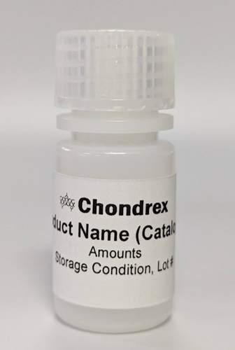
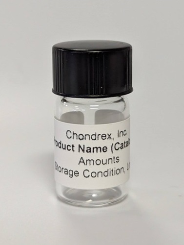
Chondrex, Inc.'s ELISA Grade Collagen is specifically prepared for use as an ELISA antigen for assays designed to quantify anti-collagen antibodies. Our ELISA Grade Collagen is native form (undenatured), monomeric collagen that has been pepsin-solubilized from various tissues. Each vial of collagen comes with 0.5 mg of collagen (enough to coat ten 96-well plates). Additionally, our 10X Collagen Dilution Buffer (Cat # 9003) is provided with each vial of ELISA Grade Collagen. This buffer is designed to prevent collagen fibril formation when coating plates. For more information, please see our recommended protocol for coating ELISA plates with ELISA Grade Collagen.
In order to accommodate the variety of collagen studies available, Chondrex, Inc. offers different grades of collagen such as immunization grade, cell culture grade, ELISA grade, T-cell assay grade, and collagen CB fragments. If you need help deciding which grade of collagen is right for your study, please see our Understanding Chondrex, Inc.'s Grades of Purified Collagen article or contact us. For more information distinguishing between Type I and Type II collagen, please see our Type I & Type II Collagen: Structure Influences Staining Patterns article or contact us.
Buffers for Collagen
| Product | Catalog # | Price (USD) | |
|---|---|---|---|
 |
Collagen Dilution Buffer, 10X concentrate, 10 ml | 9003 | 30.00 |
 |
Collagen Solubilizing Buffer | 9075 | 18.00 |
ELISA Grade Type I Collagen with 10X Buffer
| Species | Quantity | Catalog # | Price (USD) | |
|---|---|---|---|---|
 |
Bovine | 0.5 mg/ml x 1 ml | 1002 | 117.00 |
 |
Chick | 0.5 mg/ml x 1 ml | 1001 | 117.00 |
 |
Human | 0.5 mg/ml x 1 ml | 1005 | 355.00 |
 |
Mouse | 0.5 mg/ml x 1 ml | 1006 | 153.00 |
 |
Porcine | 0.5 mg/ml x 1 ml | 1003 | 117.00 |
 |
Rat | 0.5 mg/ml x 1 ml | 1004 | 154.00 |
ELISA Grade Type II Collagen with 10X Buffer
| Species | Quantity | Catalog # | Price (USD) | |
|---|---|---|---|---|
 |
Bovine | 0.5 mg/ml x 1 ml | 2012 | 222.00 |
 |
Chick | 0.5 mg/ml x 1 ml | 2011 | 222.00 |
 |
Human | 0.5 mg/ml x 1 ml | 2015 | 417.00 |
 |
Monkey | 0.5 mg/ml x 1 ml | 2017 | 269.00 |
 |
Mouse | 0.5 mg/ml x 1 ml | 2016 | 582.00 |
 |
Porcine | 0.5 mg/ml x 1 ml | 2013 | 222.00 |
 |
Rat | 0.5 mg/ml x 1 ml | 2014 | 262.00 |
ELISA Grade Type III Collagen with 10X Buffer
| Species | Quantity | Catalog # | Price (USD) | |
|---|---|---|---|---|
 |
Porcine | 0.5 mg/ml x 1 ml | 1093 | 154.00 |
Figure 1: A. Side view of an ELISA plate well coated with monomeric collagen, forming a monolayer of individual collagen molecules. B. Side view of an ELISA plate well coated with polymeric (cross-linked) collagen, creating a multi-layer of collagen proteins. This multi-layer provides multiple epitopes for anti-collagen antibodies in serum/plasma samples to bind, which can lead to an overestimation of antibody levels. Using monomeric collagen as an ELISA antigen will produce more reliable and reproducible assay results.
*Collagen shown as double helix for simplicity.
Anti-Collagen Antibodies
As a major component of connective tissues, as well as the extracellular matrix and basement membranes of many tissue types, the collagen family is the most abundant protein family in the animals. Fibril forming collagens are the most abundant types of collagen in vertebrates, but many minor collagen types have important physiological and pathological roles. In fact, the presence of antibodies against different collagen types has been associated with several autoimmune diseases:
- Anti-Type I Collagen Antibodies: Scleroderma/Systemic sclerosis (1,2)
- Anti-Type II Collagen Antibodies: Rheumatoid arthritis (RA) (3), Systemic lupus erythematosus (SLE) (4), Scleroderma/Systemic sclerosis (2)
- Anti-Type III Collagen Antibodies: Idiopathic Pulmonary Fibrosis (5)
- Anti-Type IV Collagen Antibodies: Vasculitis (6), Goodpasture Syndrome (7), Scleroderma/Systemic sclerosis (1,2)
- Anti-Type V Collagen Antibodies: Lung Allograft Rejection (8), Scleroderma/Systemic sclerosis (2)
- Anti-Type VII Collagen Antibodies: Epidermolysis bullosa acquisita (9)
However, more research is needed to determine the pathological role of anti-collagen antibodies for many of these diseases.
Anti-Collagen Antibody Assays
In addition to ELISA Grade Collagen, Chondrex, Inc. also provides ready-to-use ELISA Kits for quantifying anti-collagen antibodies from mouse, rat, and human/monkey samples. For more information on these kits, follow the links below:
- Human/Monkey Anti-Type I & II Collagen Antibody Assay Kits (IgG, IgA)
- Human Anti-Type V Collagen Antibody Assay Kits (IgG)
- Mouse Anti-Type I & II Collagen Antibody Assay Kits (IgG)
- Mouse Anti-Type I & II Collagen Antibody Subtype Assay Kits (IgG1, IgG2a, IgG2b, IgG2c, IgG3)
- Rat Anti-Type I & II Collagen Antibody Assay Kits (IgG, IgG1, IgG2a)
Diversity of Anti-Collagen Antibodies in Collagen-Induced Arthritis Models and Patients with Rheumatoid Arthritis
In the Collagen-Induced Arthritis (CIA) Model, animals receive immunizations of an emulsion comprised of heterologous type II collagen and Freund's adjuvant. CIA-susceptible animals will generate very high levels of antibodies to the heterologous collagen used for immunization. Due to the highly conserved amino acid sequence of type II collagen between species, anti-heterologous collagen antibodies in CIA-susceptible animals will cross-react to autologous type II collagen by 50% or higher. The cross reaction of antibodies to autologous type II collagen is crucial for the development of CIA. Therefore, when analyzing anti-collagen antibodies to study CIA progression, we recommend monitoring the antibody levels against the species of type II collagen used for immunization, as well as antibody levels against autologous type II collagen. This will provide the best data to correlate the development of arthritis and antibody levels.
Similarly, as humans eat products from large animals such as cow, chicken and pork, most people have antibodies to type II collagen from these species. These anti-heterologous type II collagen antibodies may cross-react to human type II collagen and could play a role in the pathogenesis of rheumatoid arthritis (RA) in particular patient groups. Furthermore, it is important to note that serum/plasma samples from humans (especially RA and other autoimmune disease patients) often have high background signals due to the non-specific binding of immunoglobulins (especially IgG) from samples to ELISA plate surfaces (9, 10). This non-specific binding is especially pronounced when sample dilution is low. Therefore, when using our ELISA Grade Collagen to assay human sera, we highly recommend using non-collagen coated plates to determine the background signal for each individual sample you plan to assay. For more information about how to account for these high background values in your assay, please contact us.
References
- Mackel AM, DeLustro F, Harper FE, LeRoy EC, Antibodies to collagen in scleroderma. Arthritis Rheum 25, 522-531, (1982).
- Riente L, et al., Anti-collagen antibodies in systemic sclerosis and in primary Raynaud's phenomenon. Clin Exp Immunol 102, 354-359, (1995).
- Mullazehi M, Wick MC, Klareskog L, van Vollenhoven R, Rönnelid J, Anti-type II collagen antibodies are associated with early radiographic destruction in rheumatoid arthritis. Arthritis Res Ther 14, R100, (2012).
- He C et al., Anti-CII antibody as a novel indicator to assess disease activity in systemic lupus erythematosus. Lupus 24, 1370-1376, (2015).
- Organ LA et al., Biomarkers of collagen synthesis predict progression in the PROFILE idiopathic pulmonary fibrosis cohort. Respir Res 20, 148, (2019).
- Wiik A, Autoantibodies in vasculitis. Arthritis Res Ther 5, 147-152, (2003).
- Cui Z et al., Antibodies to α5 chain of collagen IV are pathogenic in Goodpasture's disease. J Autoimmun 70, 1-11, (2016).
- Sumpter TL, Wilkes DS, Role of autoimmunity in organ allograft rejection: a focus on immunity to type V collagen in the pathogenesis of lung transplant rejection. Am J Physiol Lung Cell Mol Physiol 286, L1129-1139, (2004).
- Woodley DT et al., Evidence that anti-type VII collagen antibodies are pathogenic and responsible for the clinical, histological, and immunological features of epidermolysis bullosa acquisita. J Invest Dermatol 124, 958-964, (2005).
- Terato K, Do CT, Cutler D, Waritani T, Shionoya H, Preventing intense false positive and negative reactions attributed to the principle of ELISA to re-investigate antibody studies in autoimmune diseases. J Immunol Methods 407, 15-25, (2014).
- Waritani T, Chang J, McKinney B, Terato K, An ELISA protocol to improve the accuracy and reliability of serological antibody assays. MethodsX 4, 153-165, (2017).
