The extracellular matrix (ECM) is a highly dynamic and complex three-dimensional structure composed of a macromolecule network including collagen, glycosaminoglycans (GAGs), elastin, fibronectin, and several other non-cellular structures. The purpose of the ECM is to provide a homeostatic environment for cells to reside in, and although the demands for cell homeostasis differ between tissues and organs, the major constituent of ECMs is collagen (1). The collagen family of proteins is the most abundant protein family in mammals, comprising approximately 30% of total protein mass (2,3). It is the main component of connective tissues and plays a critical role in the inflammatory process of rheumatic diseases (3-5). Factors that may modulate collagen production include cancer, hypoxia, genetic disorders, inflammation, and aging (6-12). If you would like to learn more about the characteristics of collagen proteins, as well as the different grades of purified collagen Chondrex, Inc. offers, please see our Understanding Chondrex, Inc.'s Grades of Purified Collagen blog.
In order to study the physiological and pathological turnover of connective tissues and its components, Chondrex, Inc. provides Collagen Detection ELISA Kits for quantifying various species of type I and type II collagen (below), as well as total collagen detection (below), glycosaminoglycans (GAGs) detection, DNA Assay Kit, and metabolic quantification assays for tissue and cell culture samples.
To learn more about our type I collagen, type II collagen, and total collagen detection kits, please continue reading below. We provide information about the specificity of our collagen detection kits, as well as notes about preparing samples for use in our various collagen detection assays.
If you have any other questions about which collagen detection kit is right for you, please contact us.
Collagen Type I Detection Kit
| Product | Catalog # | Price (USD) | |
|---|---|---|---|
 |
Bovine Type I Collagen Detection Kit | 6014 | 519.00 |
 |
Canine Type I Collagen Detection Kit | 6019 | 519.00 |
|
Human Type I Collagen Detection Kit | 6021 | 559.00 |
|
Mouse Type I Collagen Detection Kit | 6012 | 519.00 |
|
Porcine Type I Collagen Detection Kit | 6015 | 519.00 |
|
Rabbit Type I Collagen Detection Kit | 6016 | 519.00 |
|
Rat Type I Collagen Detection Kit | 6013 | 519.00 |
 |
Type I Collagen C-Telopeptide (CTX-I) Detection Kit | 6033 | 419.00 |
|
Type I Collagen N-Telopeptide (NTX-I) Detection Kit | 6040 | 421.00 |
Collagen Type II Detection Kit
| Product | Catalog # | Price (USD) | |
|---|---|---|---|
|
Type II Collagen Detection Kit, Multi-Species | 6018 | 519.00 |
Hydroxyproline Assay Kit
| Product | Catalog # | Price (USD) | |
|---|---|---|---|
 |
Hydroxyproline Assay Kit | 6017 | 460.00 |
Reagents For Collagen Solubilization
| Product | Catalog # | Price (USD) | |
|---|---|---|---|
 |
Buffered Normal Goat Serum | 9066 | 113.00 |
 |
Elastase from Porcine Pancreas, 1 mg | 30047 | 49.00 |
 |
Elastase from Porcine Pancreas, 10 mg | 300471 | 423.00 |
 |
Normal Goat Serum | 9067 | 53.00 |
 |
Pepsin | 6011 | 15.00 |
Semi-Quantitative Tissue Total Collagen Detection
| Product | Catalog # | Price (USD) | |
|---|---|---|---|
 |
Sirius Red/Fast Green Collagen Staining Kit | 9046 | 80.00 |
 |
Sirius Red/Fast Green Dye solution, 250 ml | 90463 | 867.00 |
Sirius Red Total Collagen Detection Kit
| Product | Catalog # | Price (USD) | |
|---|---|---|---|
 |
Sirius Red Total Collagen Detection Assay PLATE Kit | 9062P | 197.00 |
 |
Sirius Red Total Collagen Detection Kit | 9062 | 143.00 |
Type I Collagen Detection ELISA Kits
Type l collagen is the most abundant collagen type and is found in most connective tissues such as skin, bone, tendon, ligament, heart, and lungs (2). A well-studied type I collagen disorder is fibrosis and the marker of lung fibrosis is an increase in collagen deposition (12). Chondrex, Inc. offers Type I Collagen Detection ELISA Kits for nine different species, including kits for C- and N-Telopeptides of type I collagen. The total assay working time is less than six hours and 40 samples can be measured in duplicate.
Species Specific Type I Collagen Detection ELISA Kits: Designed to quantify the amount of type I collagen from cell culture, tissue culture, or tissue specimens. These kits are highly specific for native form (undenatured) type I collagen, while having low reactivity to denatured collagen. Therefore, special collagen solubilization protocols must be used to prevent denaturing collagen proteins in samples. Chondrex, Inc. provides Tips for Collagen Solubilization for customers who have purchased our Type I Collagen Detection ELISA Kits. Please contact us to receive the sample preparation protocol.
Mouse and Human C-Telopeptide (CTX-I) and N-Telopeptide (NTX-I) Detection ELISA Kits: The CTX-I (competitive ELISA) and NTX-I (sandwich ELISA) Detection ELISA Kits are designed to quantify degraded type l collagen peptide fragments. Proteinases mediate resorption of type I collagen from bone and generate specific peptide fragments of degraded collagen. For example, matrix metalloproteinases (MMPs) exclusively produce C-terminal degraded fragments (ICTP) from type I collagen, while cathepsin K produces CTX-I fragments from the C-terminus and NTX-I fragments from the N-terminus of type I collagen (13). These degraded collagen fragments reflect the metabolism of type I collagen and are indicative of proteinase activities in various disease states. Therefore, immunoassays have been developed to monitor the levels of these degraded fragments in biological fluids. Chondrex, Inc. has developed CTX-I and NTX-l Detection ELISA kits for mouse and human samples using monoclonal antibodies which recognize conserved peptide sequences between the species.
Select Citations for Type I Collagen Detection Kits
Type II Collagen Detection ELISA Kit
Type II collagen is unique among the collagen family, and its tissue distribution is limited to avascular tissues such as cartilage and the vitreous body of the eyes (12,13). Because type II collagen can induce arthritis in experimental animals, autoimmunity to type II collagen is suspected in the pathogenesis of certain autoimmune diseases in humans such as rheumatoid arthritis (RA), eye diseases associated with RA, and relapsing polychondritis, which affects specific tissues containing type II collagen (13). The Type II Collagen Detection ELISA Kit is designed to quantify the amount of native type II collagen in cell/tissue cultures and tissue samples from multiple species (human, monkey, porcine, bovine, rat, mouse, rabbit, equine, and chick). The total assay working time is less than four hours and 40 samples can be measured in duplicate.
The kit is highly specific for native form (undenatured) collagen detection while having low reactivity to denatured collagen. Therefore, special collagen solubilization protocols must be used to prevent denaturing collagen proteins when preparing tissue samples for use with the assay. Chondrex, Inc. provides Tips for Collagen Solubilization for customers who have purchased our Type II Collagen Detection ELISA Kit. Please contact us to receive the sample preparation protocol.
Selected Citations for Type II Collagen Detection ELISA Kit
Hydroxyproline Assay Kit
The Hydroxyproline Assay Kit quantifies the total collagen content (any type and species) from tissue homogenates, cell cultures, and tissue culture. Hydroxyproline, a major component of collagen, makes up about 13.5% of its amino acid composition. Due to the highly restricted distribution of hydroxyproline in collagen and elastin, the hydroxyproline content generally reflects the amount of collagen in samples. Therefore, quantitating hydroxyproline has been utilized for evaluating tissue fibrosis or collagen deposition (14,15).
Conventional hydroxyproline assays require cumbersome procedures and special tools. In contrast, Chondrex, Inc.'s Hydroxyproline Assay Kit employs an improved assay system that can be operated with ease and precision using 96-well plates. The hydroxyproline assay works for determining total collagen content for both native and denatured collagen. Collagen solubilization of samples is not required, but the samples must be hydrolyzed before the assay is performed. By hydrolyzing the sample, a single hydroxyproline is formed from the total collagen sample. Please see our kit protocol to learn more about the hydrolyzation process and special considerations that need to be made. The total working time for the Hydroxyproline Assay Kit is less than one hour and 40 samples can be measured in duplicate.
Selected Citations for Hydroxyproline Assay Kit
Sirius Red/Fast Green Collagen Staining Kit- (Semi-Quantitative Assay)
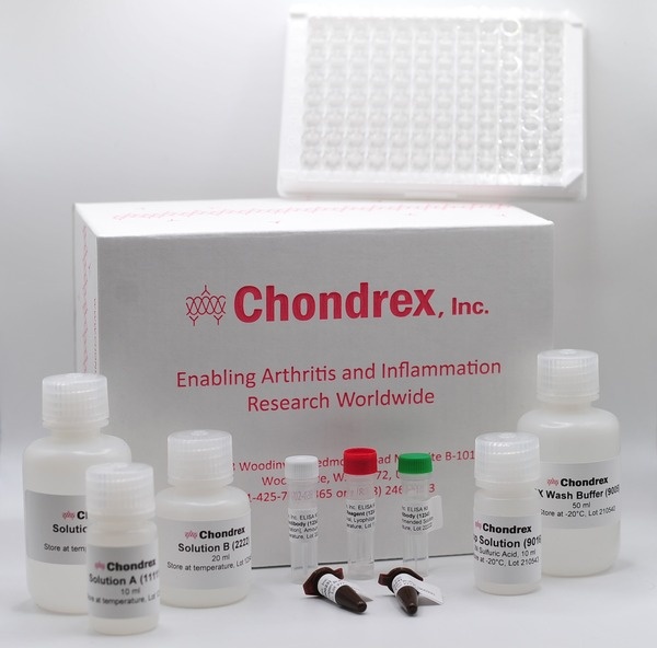
Sirius Red and Fast Green is a dye combination used to distinguish collagen from surrounding materials. Sirius Red specifically binds the [Gly-X-Y]n helical structure of fibrillar collagens, whereas Fast Green binds to non-collagenous proteins (16,17). As Sirius Red and Fast Green have absorptions at 540 nm and 605 nm respectively, the OD values of the extracted dyes can be used to calculate the collagen and non-collagenous protein content of samples. By exploiting the unique features of these two dyes, Chondrex, Inc. provides a simple semi-quantitative assay kit to determine the amounts of collagen and non-collagenous proteins in cultured cell layers and tissue sections.
Because this assay does not require collagen solubilization, it can be used to measure total collagen content in various tissues. The assay results can be normalized by results of non-collagenous proteins with Fast Green staining OD values. For general histological studies in which tissue sections are 10-20 μm thick, the assay sensitivity for collagen and non-collagenous proteins is greater than 3 μg/section and 50 μg/section, respectively. This kit contains enough reagents to stain 30-50 samples.
Selected Citations for Sirius Red/Fast Green Collagen Staining Kit
Sirius Red Total Collagen Detection Kit (Quantitative Assay)
Sirius Red Total Collagen Detection Kit utilizes Sirius red dye as a detection reagent to determine solubilized collagen concentration in samples. Sirius red is a unique dye which specifically binds to the [Gly-X-Y]n helical structure on fibrillar collagen (type I - V) (16,17). This kit does not discriminate between collagen species and types and is therefore suitable for detecting total collagen content in samples. Due to the low level of collagen in cell culture media, additional concentration steps may be necessary if assaying cell culture media samples.
The Sirius Red Total Collagen Detection Kit will only detect solubilized native (undenatured) collagen from samples. If your study requires distinguishing between native and denatured collagen, the hydroxyproline assay can be used to detect total collagen. The difference between native (Sirius red assay) and total collagen (hydroxyproline assay) will equal denatured collagen levels. While the Sirius Red Total Collagen Detection Kit is designed to be used with a spectrophotometer, our Sirius Red Collagen Detection Plate Kit is designed for use with a 96-well ELISA plate. The plate kit is convenient for assaying various collagen-containing samples (tissue specimens, cell culture media, cultured cells) at one time. The total assay working time ranges from 30-60 minutes depending on the quantity of samples and is measured at an absorption of 540 nm.
Selected Citation for Sirius Red Total Collagen Detection Kit
Extracellular matrix (ECM) assay for Glycosaminoglycans (GAGs), DNA, and MTT Assays
Besides collagen detection assays, glycosaminoglycans (GAGs) and cell proliferation assays can be used to quantify connective tissue components.
Glycosaminoglycans (GAGs) are negatively charged polysaccharides located in most connective tissues and ECM, as well as on the surfaces of many types of cell. GAGs have a widespread function and are known to play critical roles in health, hydration, and regeneration (18). Recent findings in idiopathic pulmonary fibrosis patients show an upregulation of GAGs, which possibly leads to deteriorating pro-fibrotic environment (19). In addition to type II collagen, GAGs are implicated as an autoantigen of rheumatoid arthritis (RA), as anti-GAGs antibodies associated with ECM degradation exist in serum from RA patients. In fact, immunization with GAGs can induce arthritis in mice; however, the implications of GAGs in RA pathogenesis are still under research (20,21).
Chondrex, Inc. provides a sulfated GAGs Assay Kit (cat# 6022) using the cationic dye, 1,9 dimethylmethylene blue (DMB) which binds to highly charged sulfated GAGs, except hyaluronan (21). This kit utilizes an improved DMB solution that minimizes interference with negatively charged contaminants, such as DNA and RNA. The kit also employs chondroitin sulfate as an appropriate standard for the analysis of ECM in cartilage. The total assay working time less than one hour and 40 samples can be measured in duplicate.
Deoxyribonucleic acid (DNA) and MTT (3-(4,5-dimethylthiazol2-yl)-2,5-diphenyltetrazolium bromide) assays are routinely used to assess cell proliferation and viability. These assays can be used to correlate cell health and environmental factors in culture, such as drug treatments and tissue engineering (scaffolds) (22). For example, in cartilage tissue engineering, artificial cartilage quality is evaluated by DNA amounts translated as chondrocyte numbers, as well as amounts of collagen and GAGs in the ECM (23, 24).
Chondrex, Inc. provides a DNA Assay Kit (cat# 6023) employing the Hoechst 33258 fluorescent dye which specifically binds to Adenine-Thymine base pairs, resulting in fluorescence at excitation 360 nm/emission 460 nm. As the dye-DNA binding and the fluorescence intensity are unaffected by contaminating proteins and other substances in an optimized assay condition, this DNA assay kit works accurately with samples containing other analytes (tissue samples and cell culture samples). The total assay working time is less than one hour and 40 samples can be measured in duplicate.
MTT is a positively charged compound that can enter viable eukaryotic cells and reflect cytosolic metabolic behavior under different cell culture conditions. MTT assays are routinely carried out to deduce toxicity of developed scaffolds and determine biocompatibility between collagen producing cells and scaffold materials (25). Soluble MTT is taken into cells where it is reduced, and ultimately insoluble formazan is produced and accumulates both inside and on the surface of metabolically active cells (26). The resulting formazan can be solubilized and quantified to reflect cell proliferation and viability.
Chondrex, Inc. provides an MTT Cell Proliferation Assay Kit which employs a simple method to assay cell viability and cell density using an absorbance microplate reader at 570nm. This kit can be used for up to 250 samples in duplicate.
For more details download our ECM Analysis Flow Chart Flyer and read our MTT Blog.
Selected Citations for EMC Detection Kits
Reagents for Collagen Solubilization
Both the Type I Collagen Detection Kits (species specific) and the Type II Collagen Detection Kit (multi-species) are specific for native collagen and have low reactivity with denatured collagen. Therefore, sspecial collagen solubilization protocols must be used to prevent denaturing collagen proteins in samples. Additional supplies, such as guanidine hydrochloride, pepsin (catalog # 6011, 6020), and elastase (catalog # 30047), may be necessary to process samples for use with these kits. Please contact Chondrex, Inc. to receive our Tips for Collagen Solubilization.
Buffered Normal Goat Serum (cat# 9066): Chondrex, Inc recommends pre-treatment of plastic surfaces with buffered normal goat serum to minimize non-specific binding of collagen that can lead to underestimated results. In addition, the use of blocking agent in the buffer can interfere with the results for some assays. Pre-treatment with buffered normal goat serum can minimize this risk.
Normal Goat Serum (cat# 9067): Goat serum from healthy donors used for buffer preparation.
Elastase from Porcine Pancreas (cat# 30047): Pancreatic elastase is a serine protease which will hydrolyze native elastin abundant in connective tissue. Elastase is a dissociating enzyme used for collagen solubilization from tissue samples at neutral pH. Elastase will digest cross-linkages of insoluble polymeric collagen in neutral pH tissue samples leaving a solubilized monomeric collagen as the byproduct of the reaction. Because of its effective enzymatic properties, elastase is the enzyme of choice for the isolation of Type II cells from lung tissue (27,28).
Pepsin from Porcine Gastric Mucosa (cat# 6011, 6020): The enzymatic characteristics of pepsin have been studied since 1820 and the enzyme has become widely used for countless solubilization protocols (29). Pepsin is an endopeptidase produced in the digestive system and exhibits maximum activity at pH 2.0 and is inactive at pH 6.5 and above. It can be as a dissociating enzyme for solubilizing collagen from tissue samples at an acidic pH. Pepsin cannot digest native collagen but cleaves collagen into telopeptides and atelocollagen which forms soluble collagen. However, prolonged pepsin digestion can denature collagen. Similarly, heating collagen to 40 degrees C or above can also denature collagen. Therefore, sample preparation for collagen analysis should be kept at 4 degrees C to avoid denaturation (30-32).
Selected Citation for Collagen Solubilization Reagents
References
- Theocharis AD, Skandalis SS, Gialeli C, Karamanos NK, Extracellular matrix structure. Adv Drug Deliv Rev 97:4-27, (2015). PubMed PMID: 26562801
- Ricard-Blum S, The collagen family. Cold Spring Harb Perspect Biol 3(1):a004978, (2011). PubMed PMID: 21421911
- Deshmukh SN, Dive AM, Moharil R, Munde P, Enigmatic insight into collagen. J Oral Maxillofac Pathol. 20(2):276-83, (2016). PubMed PMID: 27601823
- Rheumatoid arthritis. Nat Rev Dis Primers 4:18002, (2008). PubMed PMID: 29417950
- Fang Q, Zhou C, Nandakumar KS, Molecular and cellular pathways contributing to joint damage in rheumatoid arthritis. Mediators Inflamm 2020:3830212, (2020). PubMed PMID: 32256192
- Xu S et al., The role of collagen in cancer: from bench to bedside. J Transl Med 17(1):309, (2019). PubMed PMID: 31521169
- Ohlund D et al., Type IV collagen is a tumour stroma-derived biomarker for pancreas cancer. Br J Cancer 101(1):91-7, (2019). PubMed PMID: 19491897
- McKay TB, Hjortdal J, Priyadarsini S, Karamichos D, Acute hypoxia influences collagen and matrix metalloproteinase expression by human keratoconus cells in vitro. PLoS One 12(4):e0176017, (2017). PubMed PMID: 28426715
- Petrova V et al., The hypoxic tumor microenvironment. Oncogenesis 7:10, (2018).
- Kehlet S et al., Excessive collagen turnover products are released during colorectal cancer progression and elevated in serum from metastatic colorectal cancer patients. Sci Rep 6:30599, (2016). PubMed PMID: 27465284
- Birch HL, Extracellular matrix and ageing. Subcell Biochem 90:169-190, (2019). PubMed PMID: 30779010
- Goldstein RH, Control of type I collagen formation in the lung. Am J Physiol 261(2 Pt 1):L29-40, (1991). PubMed PMID: 1872414
- Marshall GE, Konstas AG, Lee WR, Collagens in ocular tissues. Br J Ophthalmol 77(8):515-524, (1993). PubMed PMID: 8025051
- Cremer MA, Pitcock JA, Stuart JM, Kang AH, Townes AS, Auricular chondritis in rats. An experimental model of relapsing polychondritis induced with type II collagen. J Exp Med 154(2):535-40, (1981). PubMed PMID: 7021752
- Wheater G et al., The clinical utility of bone marker measurements in osteoporosis. J Transl Med 11:201, (2013). PubMed PMID: 23984630
- Cundy T, Reid IR, Grey A, CHAPTER 31 - Metabolic bone disease. Clinical Biochemistry: Metabolic and Clinical Aspects (Churchill Livingstone, Third Edition, 2014) Pages 604-635, ISBN 9780702051401.
- Segnani C et al., Histochemical detection of collagen fibers by sirius red/fast green is more sensitive than van Gieson or sirius red alone in normal and inflamed rat colon. PLoS One 10(12):e0144630, (2015). PubMed PMID: 26673752
- Casale J, Crane JS, Biochemistry, glycosaminoglycans. Treasure Island (FL): StatPearls Publishing. (2020).
- Westergren-Thorsson G et al., Increased deposition of glycosaminoglycans and altered structure of heparan sulfate in idiopathic pulmonary fibrosis. The international journal of biochemistry & cell biology 83:27-38, (2017). PubMed PMID: 27974233
- Wang JY, Roehrl MH, Glycosaminoglycans are a potential cause of rheumatoid arthritis. Proc Natl Acad Sci USA. 99(22):14362-7, (2002). PubMed PMID: 12391302
- Barbosa I et al., Improved and simple micro assay for sulfated glycosaminoglycans quantification in biological extracts and its use in skin and muscle tissue studies. Glycobiology 13:647-653, (2003). PubMed PMID: 12773478
- Pabbruwe MB et al., A comparison of colorimetric and DNA quantification assays for the assessment of meniscal fibrochondrocyte proliferation in microcarrier culture. Biotechnology letters 27(19):1451-5, (2005). PubMed PMID: 16231215
- Yoon JH, Halper J, Tendon proteoglycans: biochemistry and function. J Musculoskelet Neuronal Interact 5:22-34 (2005). PubMed PMID: 15788868
- Buckwalter JA, Mankin HJ, Articular cartilage: tissue design and chondrocyte-matrix interactions. Instr Course Lect 47:477-486, (1998). PubMed PMID: 9571449
- Varkey A et al., Impact of silk fibroin-based scaffold structures on human osteoblast MG63 cell attachment and proliferation. Int J Nanomedicine 10 Suppl 1(Suppl 1):43-51, (2015). PubMed PMID: 26491306
- Twentyman PR, Luscombe M, A study of some variables in a tetrazolium dye (MTT) based assay for cell growth and chemosensitivity. Br J Cancer 56:279-285 (1987). PubMed PMID: 3663476
- Worthington Tissue Dissociation Guide by Worthington Biochemical Corporation http://www.worthington-biochem.com/tissuedissociation/elastase.html
- Fields GB, Interstitial collagen catabolism. J Biol Chem 288(13):8785-93, (2013). PubMed PMID: 23430258
- Last JA, Reiser KM, Collagen biosynthesis. Environ Health Perspect 55:169-77, (1984). Review. PubMed PMID: 6428877
- Fruton JS, A history of pepsin and related enzymes. Q Rev Biol 77(2):127-47, (2002). Review. PubMed PMID: 12089768
- Johnston N, Dettmar PW, Bishwokarma B, Lively MO, Koufman JA, Activity/stability of human pepsin: implications for reflux attributed laryngeal disease. Laryngoscope 117(6):1036-1039, (2007). PubMed PMID: 17417109
- Dunn BM, Overview of pepsin-like aspartic peptidases. Current Protocols in Protein Science Chapter 21 (1): 21.3.1-21.3.6, (2001) PubMed PMID: 18429164